Home
/ Major Muscles In The Body Diagram / Pin by ashlee brown on Anatomy | Leg muscles anatomy ... - Located immediately below the skin) it should be noted that there are many more muscles in the body that are not addressed by this muscle anatomy diagram, however the muscles that are of primary.
Major Muscles In The Body Diagram / Pin by ashlee brown on Anatomy | Leg muscles anatomy ... - Located immediately below the skin) it should be noted that there are many more muscles in the body that are not addressed by this muscle anatomy diagram, however the muscles that are of primary.
Major Muscles In The Body Diagram / Pin by ashlee brown on Anatomy | Leg muscles anatomy ... - Located immediately below the skin) it should be noted that there are many more muscles in the body that are not addressed by this muscle anatomy diagram, however the muscles that are of primary.. There are around 650 skeletal muscles within the typical human body. It is formally known as the triceps brachii muscle. The human muscular system is complex and has many functions in the body. Both are supremely tough, which you'll quickly realise. See how all sharpness disappears?
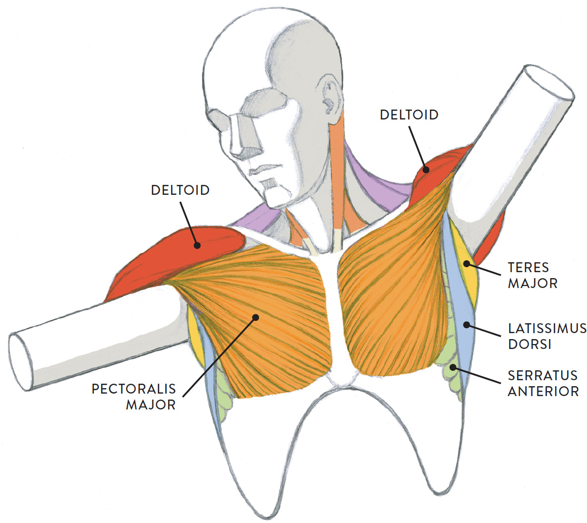
Anterior muscles in the body. A chest muscle that pulls the arm in towards the body. It is formally known as the triceps brachii muscle. Despite their similar names, teres major has different actions and innervation from the teres minor. Skeletal muscles produce movements of the skeleton and other body parts.
What are Muscles | What do Muscles do | DK Find Out from res.cloudinary.com √ describe how muscles work in pairs to make parts. The thigh adductors pull the legs together when. These muscles are the only voluntary muscles in the body—we can control these muscles. This is what happens in the body. Human muscle system, the muscles of the human body that work the skeletal system, that are under voluntary control, and that are concerned the quadratus lumborum muscle in the lower back side bends the lumbar spine and aids in the inspiration of air through its stabilizing affects at its insertion at. Their main function is contractibility. This set is often saved in the same folder as. Click on the labels, or move the mouse over the diagram to see the muscles identified.
Teres major is a thick and ovoid muscle in the upper arm.
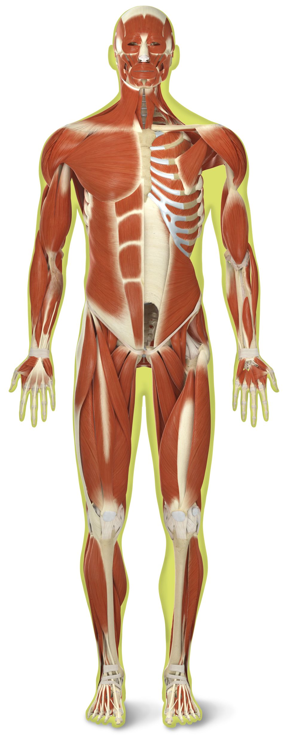
Major muscles of the human body. This large muscle in the back of the upper arm helps straighten the arm. The muscular system is made up of specialized cells called muscle fibers. √ describe how muscles work in pairs to make parts. Strongest and leanest of all muscles in the body. Their main function is contractibility. Despite their similar names, teres major has different actions and innervation from the teres minor. The psoas major may hold the distinction of being the most important muscle in the human body and at the same the most misunderstood muscle in note: Click on the labels, or move the mouse over the diagram to see the muscles identified. The interactive muscle anatomy diagram shown below outlines the major superficial (i.e. Located immediately below the skin) it should be noted that there are many more muscles in the body that are not addressed by this muscle anatomy diagram, however the muscles that are of primary. These muscles hold the inner ear together and are connected to. **** sorry i made a mistake at 00:49 i incorrectly label and describe the thigh adductors as hip abductors.
In the muscular system, muscle tissue is categorized into three distinct types: **** sorry i made a mistake at 00:49 i incorrectly label and describe the thigh adductors as hip abductors. See how all sharpness disappears? Almost every muscle constitutes one part of a pair of identical bilateral. Today we'll be looking at the 10 largest pectoralis major.
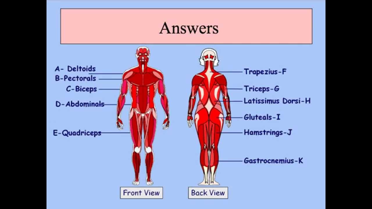
MUSCLE DIAGRAM from schoolbag.info This quiz requires labeling, so it will test your knowledge on how to identify these muscles (latissimus dorsi, trapezius, deltoid, biceps brachii. This diagram with labels depicts and explains the details of major muscles in the human body. Teres major is a thick and ovoid muscle in the upper arm. Human muscle system, the muscles of the human body that work the skeletal system, that are under voluntary control, and that are concerned the quadratus lumborum muscle in the lower back side bends the lumbar spine and aids in the inspiration of air through its stabilizing affects at its insertion at. Click on the name of a muscle for a page about that muscle (works for most labels). Each workout involves working through three supersets, and both target all the major muscle groups in the back. The interactive muscle anatomy diagram shown below outlines the major superficial (i.e. Some cardiac muscles are also present in the walls of the aorta.
See how all sharpness disappears?
Diagram of the human body. This muscle diagram is interactive: Human muscle system, the muscles of the human body that work the skeletal system, that are under voluntary control, and that are concerned the quadratus lumborum muscle in the lower back side bends the lumbar spine and aids in the inspiration of air through its stabilizing affects at its insertion at. This article is modified from an article originally published in the massage therapy journal (mtj): The human muscular system is complex and has many functions in the body. A chest muscle that pulls the arm in towards the body. Chart of major muscles on the front of the body with labels. This large muscle in the back of the upper arm helps straighten the arm. If you found any images copyrighted to yours, please contact us and we. Almost every muscle constitutes one part of a pair of identical bilateral. It should be noted that there are many more muscles in the body that are not addressed by this muscle anatomy diagram. Muscle anatomy quiz for anatomy and physiology! Smooth muscle contractions are involuntary movements triggered by.
When you are taking anatomy and physiology you will be required to identify major muscles in the human body. The size of the muscle can be used to muscles are the only tissue in the body that has the ability to contract and therefore move the. The thigh adductors pull the legs together when. These include mobility, stability, posture, circulation, digestion, and more. It is a perfect combination of multiple muscles working in harmony and.
GCSE PE Podcast Muscular system - YouTube from i.ytimg.com Located in the front of your upper arm, large muscle in the back upper part of the arm, muscle located at the top of the forearm, muscle next to brachioradialis. Strongest and leanest of all muscles in the body. There are around 650 skeletal muscles within the typical human body. We hope this post inspired you and help you what you are looking for. It joins the iliacus muscle to form the iliopsoas. Located immediately below the skin) muscles of the body. This is a table of skeletal muscles of the human anatomy. Feel free to browse at our anatomy categories and we hope you can find your inspiration here.
Skeletal muscles produce movements of the skeleton and other body parts.
The size of the muscle can be used to muscles are the only tissue in the body that has the ability to contract and therefore move the. Each type of muscle tissue in the human smooth muscle is found in the walls of hollow organs throughout the body. This set is often saved in the same folder as. The thigh adductors pull the legs together when. Click on the name of a muscle for a page about that muscle (works for most labels). Click on the labels, or move the mouse over the diagram to see the muscles identified. Located in the front of your upper arm, large muscle in the back upper part of the arm, muscle located at the top of the forearm, muscle next to brachioradialis. Today we'll be looking at the 10 largest pectoralis major. **** sorry i made a mistake at 00:49 i incorrectly label and describe the thigh adductors as hip abductors. Thank you for visiting major muscles of the body diagram pictures. In the muscular system, muscle tissue is categorized into three distinct types: There are around 650 skeletal muscles within the typical human body. Skeletal muscles produce movements of the skeleton and other body parts.
Anterior muscles in the body muscles in the body diagram. Some cardiac muscles are also present in the walls of the aorta.